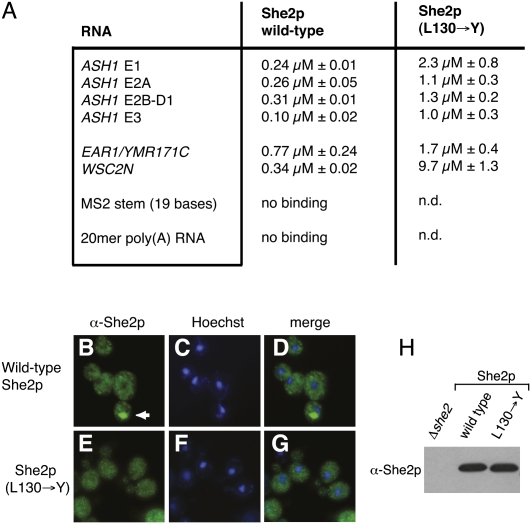
FIGURE 6.
Functional analyses with wild-type and oligomerization-mutant She2p. (A) RNA filter-binding assays with She2p. All Kd values shown are derived from at least three independent binding experiments. The label “no binding” indicates that at the highest protein concentration measured, no significant RNA binding was observed (see Materials and Methods). “n.d.” indicates that an experiment has not been performed. (B–G) Bud–tip localization in response to mutation of amino acid 130 in She2p. (B–D) Antibody staining reveals that wild-type She2p localizes to the bud tip in 69.2% of otherwise ΔSHE2 cells. (E–G) In contrast, no localization above background level (i.e., 3%) was observed in ΔSHE2 cells expressing She2p (L130→Y). (B,E) show anti-She2p antibody staining, (C,F) show nuclear Hoechst staining, and (D,G) display merged images of the respective antibody and nuclear stainings. Arrow indicates the position of She2p localization at the bud tip. In all experiments, at least 100 budding cells were inspected for She2p localization. (H) Western-blot analysis with anti-She2p antibody shows that She2p (L130→Y) is expressed at wild-type levels.

