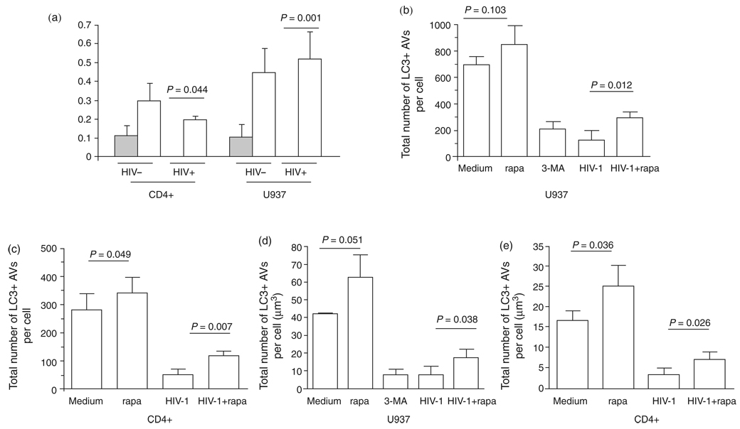
Fig. 2. Induction of autophagy. Cells were incubated for 48 h following treatment with media only or infectious HIV-1 and then half of the cells were starved for 3 h.
(a) Beclin 1 protein expression levels of the cells undergoing starvation are represented by open bars and those without starvation by shaded bars. Beclin 1 and GAPDH proteins were probed by western blot. (b–e) Autophagosome number and volume were detected by fluorescent LC3 staining after autophagy induction. Results are representative of three independent experiments.

