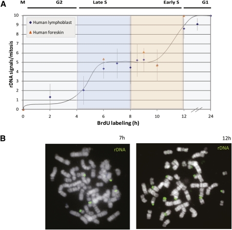
Figure 2.
Replication timing of rDNA. (A) Human lymphoblasts or foreskin fibroblasts were labeled with BrdU for various times, arrested in metaphase, and subjected to ReTiSH using a probe for rDNA. The number of rDNA signals in each metaphase spread (n = 461) was counted, and standard deviation was calculated for each data point (see error bars). (B) Examples at 7 and 12 h are shown.

