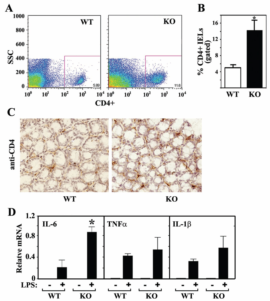
Figure 3. CD4+ IELs are upregulated in colons of Foxo4 KO mice.
(A and B) Relative percentages of CD4+ IELs from colons of WT and Foxo4-KO mice were analyzed by flow cytometry on the basis of forward/side scatter-based lymphocyte gating (n=3 ± SEM, *, p<0.05). (C) Colonic CD4+ IELs were stained with anti-CD4 antibody (dark brown) in an immunohistochemistry assay. A representative IHC staining from three different mice per genotype is shown. (D) Relative mRNA in peritoneal macrophages stimulated with or without LPS (n=3 ± SEM, *, p<0.05).

