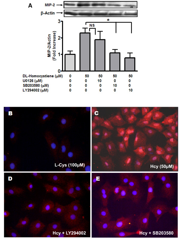
Figure 2.
Homocysteine-induced MIP- 2 is mediated by p38MAPK and PI3 kinase. MCs were incubated (24 hours; 37°C) in the presence of Hcy (50 μM) with or without inhibitors U0126 (p42/44 MAPK inhibitor; 10 μM), SB203580 (p38MAPK inhibitor; 10 μM) and LY294002 (PI3 Kinase inhibitor; 10 μM). Cells were washed with PBS (4°C) and harvested using lysis buffer under non-denaturing conditions. MIP-2 protein was detected by western blot (A). Subsequently, protein bands were quantified as before. Results are representative of three separate experiments. Data represent mean ± SEM; *p < 0.05 indicate significant inhibition compared to 50 μM Hcy. (B to E) MCs were incubated (24 hours; 37°C) in the presence of Hcy (50 μM) with or without kinase inhibitors in Lab-Tek II dual chamber slides (Nalge Nunc, Naperville, IL, USA). The fixed MCs were immuno-stained with rabbit polyclonal GRO beta antibody followed by Alexa-Fluor 555 conjugated anti-rabbit antibody as described in the method. Nuclei were stained with DAPI. Panel B: L-Cys [100 μM], Panel C: Hcy [50 μM]; Panel D: Hcy [50 μM] + LY294002 [10 μM]; Panel E: Hcy [50 μM] + SB203580 [10 μM]. Panels are representative of 3 separate experiments.

