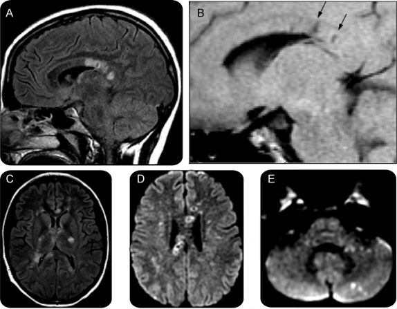
Figure 1 MRI on the day of presentation
(A) T2 fluid-attenuated inversion recovery (FLAIR) parasagittal view shows corpus callosal lesions, some with a ring of increased signal and a darker center. (B) T1 parasagittal view, similar cut, shows areas of T1 hypointensity (arrows) corresponding to the T2 bright lesions. (C) T2 FLAIR axial views show additional lesions throughout the internal capsule, and the genu, splenium, and tapetum of the corpus callosum. (D) Diffusion-weighted imaging (DWI) (1000b) axial view through the superior extent of the lateral ventricles shows several lesions with restricted diffusion through the central fibers of the corpus callosum, many with bright rings and dark centers. (E) DWI (1000b) axial view of the cerebellum and pons shows pinpoint lesions in the middle cerebellar peduncle and cerebellar cortex.

