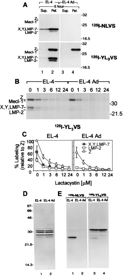
Figure 1.
Labeling of subcellular fractions and purified proteasomes from EL-4 and adapted cells with covalent active site-directed affinity probes. (A) Twenty micrograms of each subcellular fraction was incubated with 125I-NLVS or 125I-YL3VS for 2 h at 37°C, resolved by 12.5% SDS/PAGE, and visualized by autoradiography. The affinity probe labeling of proteasomal β subunits labeled is described elsewhere (23). (B) Lactacystin was incubated with intact EL-4 and adapted EL-4 cells for 12 h at 37°C, lysed, labeled with 125I-YL3VS, and analyzed by 12.5% SDS/PAGE and autoradiography. (C) Quantitative analysis of results in Fig. 3B. Bands representing the Z, X/Y/LMP-7, or LMP-2 subunits were quantified by using PhosphorImager analysis (imagequant, Molecular Dynamics). Mean values and standard deviations of three separate experiments are shown. (D) Purified 20S from EL-4 and adapted cells were incubated with 125I-NLVS or 125I-YL3VS for 2 h at 37°C and visualized by silver staining the 12.5% SDS/PAGE and by autoradiography (E). Note: in proteasomes from adapted cells, LMP-7 is modified by NLVS and comigrates with LMP-8 (A, lane 2) (18).

