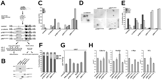
Figure 2. Effect of the t(9;22) fusion proteins on the biology of murine HSCs.
(A) Experimental strategy for studying the influence of the t(9;22) fusion proteins on the biology of murine HSCs. Sca1+/lin− BM cells were infected with the indicated retroviruses and plated in semi-solid medium supplemented with the indicated growth factors for determination of the serial replating potential. Cells from the first plating (I) round were examined for the expression of differentiation-specific surface markers. Cells plated in liquid culture supplemented with the indicated growth factors were used for cell cycle analysis and gene expression by qRT-PCR. (B) Sca1+/lin− BM cells were infected with the indicated retroviruses and expression levels of the transgenes were analyzed by Western blotting using the indicated antibodies. (C) Long-term serial replating. Sca1+/lin− cells were infected with the indicated retroviruses and plated into methyl-cellulose supplemented with the indicated growth factors to assess primary colony formation. Colony numbers were counted on days 8–10. Cells were then harvested and serially replated. Colonies were counted on days 8–10 after each replating. (D) Colony morphology during the first plating. Type A (compact colonies), type B (dense center surrounded by a halo of migrating cells) and type C (diffuse colonies with mobile differentiating cells) colonies were distinguished. (E) Expression of differentiation-specific surface markers. One representative experiment of three yielding similar results. (F) Sca1+/lin− BM cells were infected with the indicated retroviruses and, after 48 h, cell cycle progression was determined. The provided results are the average of three independent experiments +/− SD. (G) Activation of Wnt-signaling by the t(9;22) fusion proteins. BCR, p185BCR/ABL, p40ABL/BCR and p96ABL/BCR and the Topflash and Fopflash reporter constructs were co-transfected by electroporation into U937 cells. U937 cells expressing PML/RARα - positive control; pGL3basic - negative control. Luciferase activity measured at 24 h post-transfection was normalized to Renilla luciferase activity. Each experiment was performed in triplicate a total of three times with similar results. One representative experiment is given +/− SD. (H) Sca1+/lin− BM cells were infected with the indicated retroviruses and, at 48 h post-infection, the expression levels of HoxB4, Cdkn1a (p21(cip1/waf1)), c-Myc and SCL were analyzed by qRT-PCR. The relative concentration of each mRNA was normalized to the concentration of the housekeeping gene GAPDH and is represented as 2−Δ/Δ CT. Each experiment was performed in triplicate a total of three times with similar results. One representative experiment is given +/− SD.

