Fig. 2.
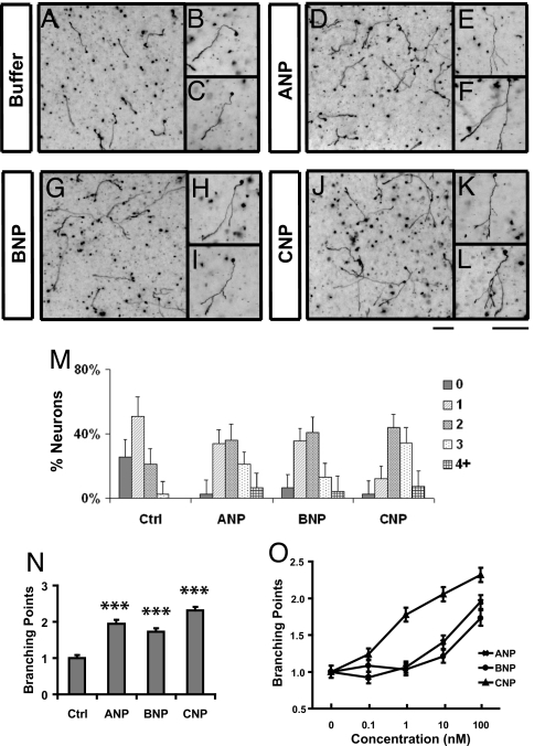
NPs promote axon branching of dissociated DRG neurons in culture. (A–L) Dissociated E14 DRG neurons were cultured in collagen gels in the presence of NGF (25 ng/mL) for 24 h and then treated with buffer (control, A–C) or 100 nM ANP (D–F), BNP (G–I), or CNP (J–L). After being cultured for another day, they were fixed and stained with neurofilament antibodies. Regions of the cultures are shown at low magnification in A, D, G, and J, whereas individual cells are shown at high magnification in B, C, E, F, H, I, K, and L. Neurons treated with all NPs showed a significant increase in branch formation. (Scale bars: 100 μm.) (M and N) Distribution of neurons with different numbers of branches (M) and the average number of branching points (N) (***, P < 0.001, ANOVA test) measured from the above cultures treated with different NPs (100 nM). (O) Comparison of the average number of branching points in the cultures treated with different doses of NPs. More than 80 neurons were analyzed for each condition, and the results were plotted as mean ± SD in M and mean ± SEM in N and O.

