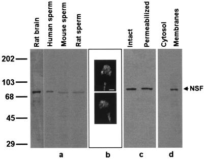
Figure 1.
NSF is present in membranes of human spermatozoa and localizes to the acrosomal region. (a) Proteins from rat brain and human, mouse, and rat sperm were extracted in Laemmli sample buffer and analyzed by Western blot using an anti-NSF mAb as probe. Lanes: rat brain, 1 μg of a postnuclear membrane pellet from rat brain; human sperm, proteins from 5 × 10⁶ cells; mouse sperm, proteins from 10 × 106 cells; rat sperm, proteins from 4 × 10⁶ cells. Molecular mass standards (kDa × 10−3) are indicated to the left. (b) Human sperm were fixed and permeabilized and immunostained with a rabbit polyclonal anti-NSF antibody followed with a TRITC-labeled goat anti-rabbit antibody. Shown are epifluorescence micrographs of two typically stained cells. (Bar = 2 μm.) (c) Proteins extracted in 1% Triton X-100 from intact (intact lane) or SLO-permeabilized (permeabilized lane) were denatured in Laemmli sample buffer and analyzed by Western blot using an anti-NSF mAb as probe (7 × 10⁶ cells per lane). (d) Percoll-washed human sperm were separated into soluble (cytosol lane) and particulate (membranes lane) fractions and probed on blots with an anti-NSF mAb. Ten micrograms of proteins derived from 1.7 × 10⁸ cells was loaded per lane.

