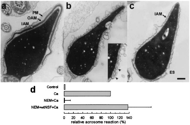
Figure 3.
Morphological observation of acrosomal exocytosis in permeabilized spermatozoa. The acrosome reaction was evaluated by transmission electron microscopy in permeabilized spermatozoa after incubation under the following conditions: Control, no additions; Ca, 0.5 mM CaCl2 in the presence of 0.5 mM EGTA; NEM+Ca, spermatozoa were treated with 0.6 mM NEM during permeabilization and stimulated with calcium; NEM+wtNSF+Ca, pretreatment with 0.6 mM NEM and addition of 300 nM wild-type NSF and calcium. At the end of the incubation, the samples were fixed in 2% glutaraldehyde and analyzed by transmission electron microscopy. Examples of unreacted and reacted spermatozoa are shown in a–c. The unreacted sperm (NEM+Ca) in a present intact acrosomal granule and plasma membrane (PM, plasma membrane; OAM, outer acrosomal membrane; IAM, inner acrosomal membrane). In b, a vesiculated spermatozoon (NEM+NSF+Ca), presumptively undergoing acrosomal exocytosis, is shown. Notice the multiple interactions between the outer acrosomal and plasma membrane, particularly evident in the equatorial segment enlarged in the Inset (arrowheads). (c) A completely reacted spermatozoon (NEM+NSF+Ca). Only the IAM is observed above the equatorial segment (ES). (Bar = 0.5 μm.) (d) The percentage of spermatozoa lacking acrosomes was evaluated for each condition and normalized as explained in Material and Methods (ranges for negative and positive controls were 10–13% and 20–29%, respectively). The data represent the means and ranges of two independent experiments.

