
Figure 3 (A–B).
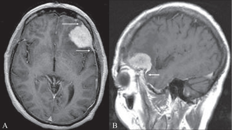
Dural tail sign. Axial (A) and sagittal (B) contrast-enhanced MRI images of the brain show an avidly enhancing lesion in the left frontal region. The lesion is dural based and there is thickening and enhancement of the dura, which tapers away from the mass (arrows). This thickening and enhancement of the dura adjacent to the mass is the ‘dural tail sign’ that is seen in meningiomas