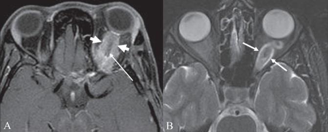
Figure 10 (A, B).
Optic nerve tram-track sign. Postcontrast T1W axial MRI image of the orbits (A) shows an enhancing mass (thick small arrows) around the left optic nerve. The optic nerve (thin long arrow) itself is not enlarged and is nonenhancing. This is suggestive of a mass, such as a meningioma, arising from the optic nerve sheath. Axial T2W MRI image of the orbits in a different patient (B) shows fusiform enlargement of the left optic nerve itself (arrows), in a case of optic nerve glioma

