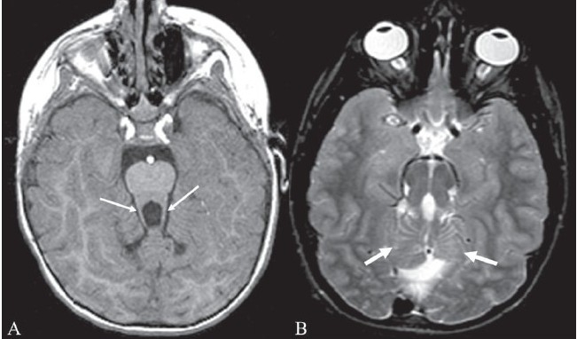
Figure 15 (A, B).

Molar tooth sign. Axial T1W MRI image of the brain (A) in a child with Joubert syndrome shows elongated superior cerebellar peduncles (arrows) giving the midbrain and superior peduncles the appearance of a molar tooth. Axial T2W image of the brain (B) in another child shows the molar tooth appearance of the midbrain and superior cerebellar peduncles. There is also hypoplasia of the cerebellar vermis (arrows)
