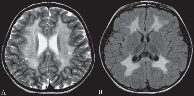
Figure 16 (A, B).

Tigroid pattern. Axial T2W image of the brain in a child with metachromatic leukodystrophy shows symmetric, increased signal intensity of the white matter, with sparing of the subcortical U fibers. Linear low signal intensity areas radiating away from the ventricular margin (arrows) represent areas of white matter around the vessels that have been spared from the process of demyelination. These low signal linear areas within the hyperintense white matter resemble the skin of a leopard and hence the term ‘tigroid’ pattern. FLAIR axial MRI image (B) shows symmetric hyperintensity of the white matter sparing the subcortical U fibers
