
Figure 17 (A, B).
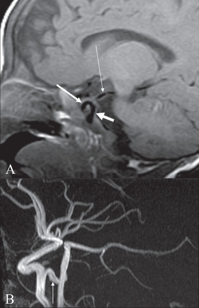
Tau sign. Sagittal T1-W image of the brain shows (A) flow voids of the internal carotid artery (ICA) in the precavernous segment (thick short arrow), in the cavernous segment (medium-sized arrow), and a persistent trigeminal artery (thin long arrow). Together, these flow voids form the Greek letter ‘τ’ (tau). Sagittal view of the MRI angiogram shows the persistent trigeminal artery (arrow) arising from the ICA and joining the basilar artery in its mid segment