
Figure 1 (A-C).
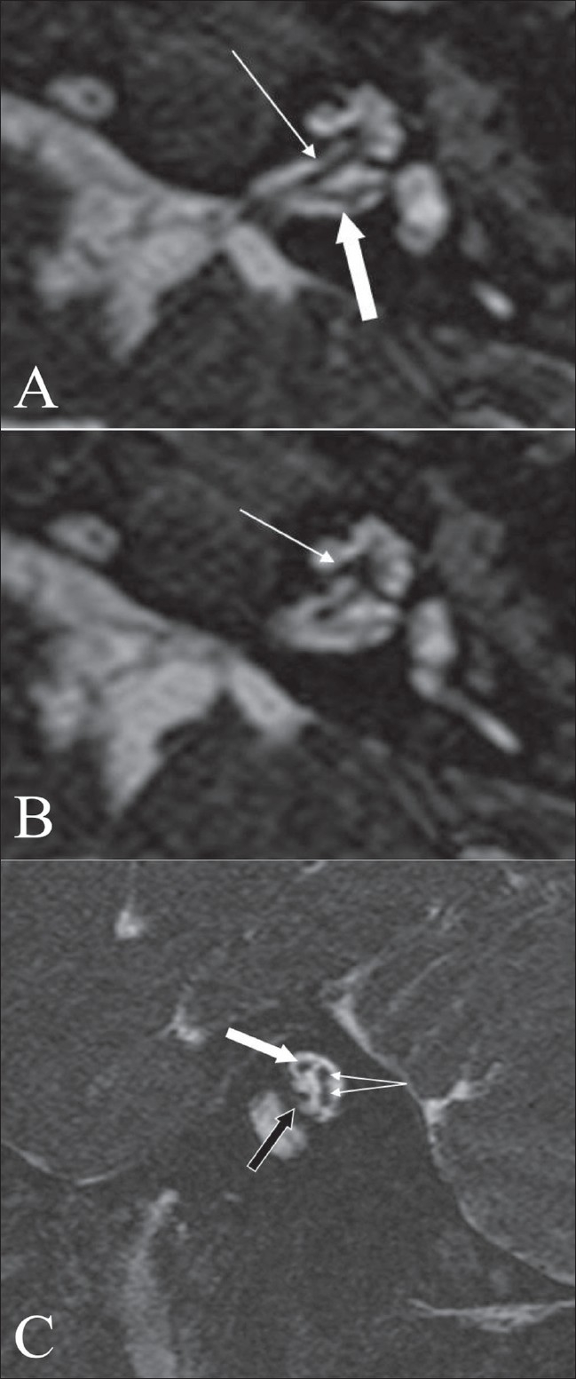
Normal anatomy. An axial 3D-FIESTA image (A), at the level of the vestibule and cochlea shows the cochlear (arrow) and vestibular (block arrow) nerves. An axial 3D-FIESTA image (B), at the level of the vestibule and cochlea, shows a well-delineated and intact modiolus (arrow). An oblique sagittal 3D FIESTA image (C) through the internal auditory canal shows the facial nerve in the antero-superior quadrant (block white arrow), the cochlear nerve in the antero-inferior quadrant (block black arrow) and the superior and inferior vestibular nerves, in the posterior quadrant (branching arrows)