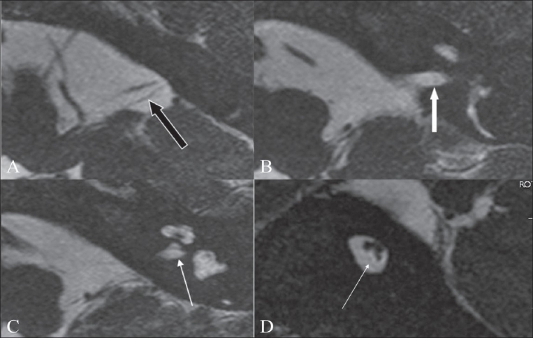
Figure 9 (A-D).

Cochlear nerve deficiency. Axial 3D-FIESTA images (A-C) show a markedly thinned-out eighth nerve in the CP angle cistern (black arrow in A), which is barely seen in the distal portion of the canal (white arrow in B). The left cochlear nerve is not visualized in its normal location (arrow in C). An oblique sagittal 3D-FIESTA image (D) shows the facial and vestibular nerves in their normal location, with the cochlear nerve barely or almost not seen in the antero-inferior quadrant (arrow)
