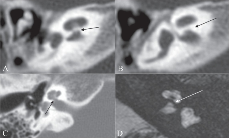
Figure 10 (A-D).

Isolated cochlea. Axial HRCT images (A, B) show a thick bony bar at the cochlear aperture (arrow), suggesting an isolated cochlea. An axial HRCT image (C) shows a normal appearance of the cochlear aperture at the site of the entry of the cochlear nerve, near the modiolus. An axial 3D-FIESTA image (D) shows a bony bar (arrow) at the cochlear aperture (isolated cochlea). Also note the left cochlear nerve deficiency
