Figure 2.

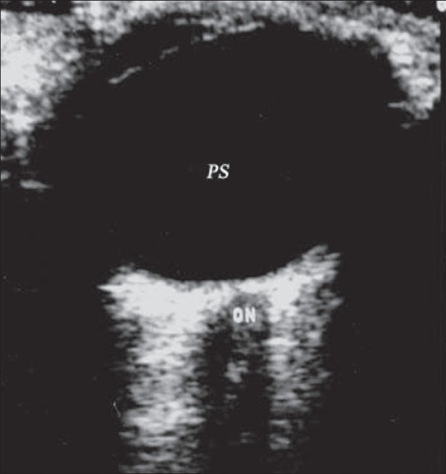
Normal anatomy. B-scan reveals a normal eyeball with the optic nerve (ON) passing through the retrobulbar fat. Normal posterior segment (PS) contains clear anechoic vitreous

Normal anatomy. B-scan reveals a normal eyeball with the optic nerve (ON) passing through the retrobulbar fat. Normal posterior segment (PS) contains clear anechoic vitreous