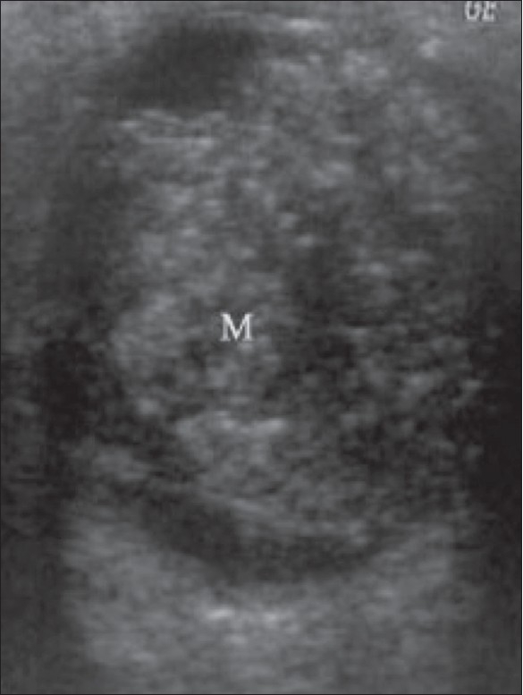
Figure 14.

Retinoblastoma. This is the case of a child with leukocoria. B-scan reveals a hyperechoic tumor (M) extensively filling the posterior segment. The calcium deposits, seen as highly reflective foci, are pathognomonic. The tumor outline is irregular
