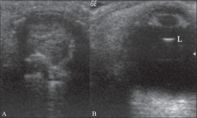
Figure 16 (A,B).

Phthisis. The left eyeball is clinically blind. The B-scan of the left eye (A) shows a shrunken globe with extensive calcification and loss of the normal shape. There was a history of trauma. The normal right eyeball (B) is shown for comparison
