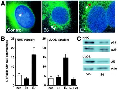
Figure 2.
(A) NHKs were transiently transfected with HPV-16 E6 or E7 and a vector encoding farnesylatable GFP. Only GFP-positive cells were evaluated. Keratinocytes transfected with the parental plasmid were used as the control. Centrosomes were visualized by immunofluorescence staining for γ-tubulin (arrowheads). Nuclei were counterstained with the DNA dye Hoechst 33258. Pictures were obtained with a multiband filter set. (B) Quantitation of centrosomal abnormalities in NHKs and U2OS cells transiently transfected with HPV-16 E6 or E7. Cells transfected with the parental plasmid (neo) were used as the control. Bar graphs show the mean ± SEM of three independent experiments. (C) Decreased p53 levels in response to transient HPV-16 E6 expression in NHKs (Upper) and U2OS cells (Lower). An actin blot is shown to demonstrate equal loading.

