Figure 7.
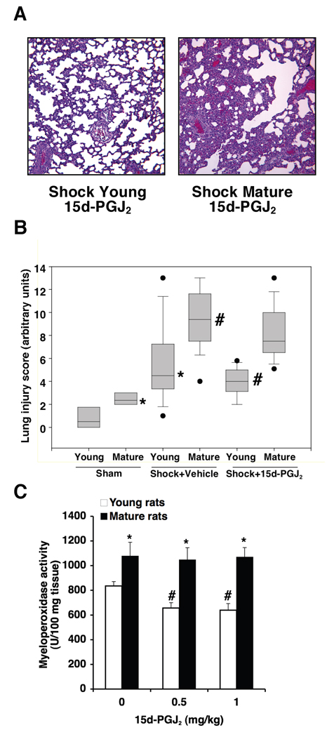
(A) Representative lung histology of a young and a mature rat subjected to hemorrhage (3 h) and resuscitation (3 h) and treated with 15d-PGJ2 (0.5 mg/kg i.p.). For comparison with vehicle-treated rats and sham groups see Figure 2. Magnification x 100. A similar pattern was seen in n=10 lung sections in each experimental group. (B) Histopathological scores of lung sections (of n=5–10 rats for each group). Lung injury was scored from 0 (no damage) to 16 (maximum damage). Box plots represent 25th percentile, median and 75th percentile; error bars define 10th and 90th percentile; black dots define outliers. *Represents p < .05 versus young sham rats. #Represents p < .05 versus young vehicle-treated rats subjected to hemorrhagic shock. Treated rats received 15d-PGJ2 (0.5 mg/kg i.p.). (C) Effect of treatment with 15d-PGJ2 (0.5–1 mg/kg i.p.) on lung myeloperoxidase activity in young and mature rats subjected to hemorrhage (3 h) and resuscitation (3 h). Each data point represents the mean ± SEM of 5–16 rats for each group. *Represents p < .05 versus young rats. #Represents p < .05 versus vehicle-treated rats (i.e., 0 mg/kg).

