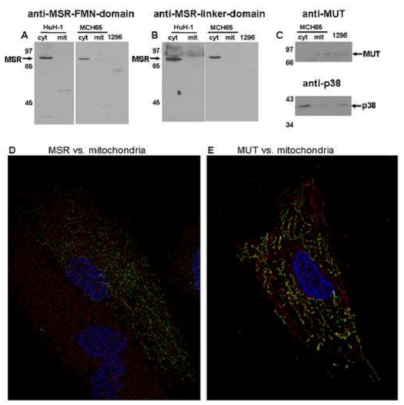
Figure 4. Subcellular localization of MSR. Panels A & B.

Western blot of cytosolic (cyt) and mitochondrial (mit) fractions of Huh-1 and MCH65 cells and total cell protein of WG1296 (1296) probed with anti-MSR-FMN-domain (panel A) or anti-MSR-linker-domain (panel B). Panel C. Control to test purity of mitochondrial and cytoplasmic fractions. Upper panel shows anti-MUT (mitochondrial protein); lower panel shows anti-p38 (cytosolic and nuclear protein). There appears to be a small amount of cytosolic contamination of the mitochondrial fraction (band in ‘mit’ of p38), but almost no mitochondrial contamination of cytoslic fraction (band ‘c’ in MUT). Lanes are the same as indicated for Panel A and B. Panel D & E. Immunofluorescent localization of MSR. MCH65 cells were transfected with pAcGFP1-mito (mitochondrial label, Green) and were immunostained for either MSR (anti-MSR-linker-domain) Panel D or methylmalonyl-CoA mutase (anti-MUT) Panel E (Red). Nuclei are stained with DAPI (Blue). After deconvolution, MUT was found to co-localize with mitochondria (yellow) but MSR was not.
