
Figure 1.
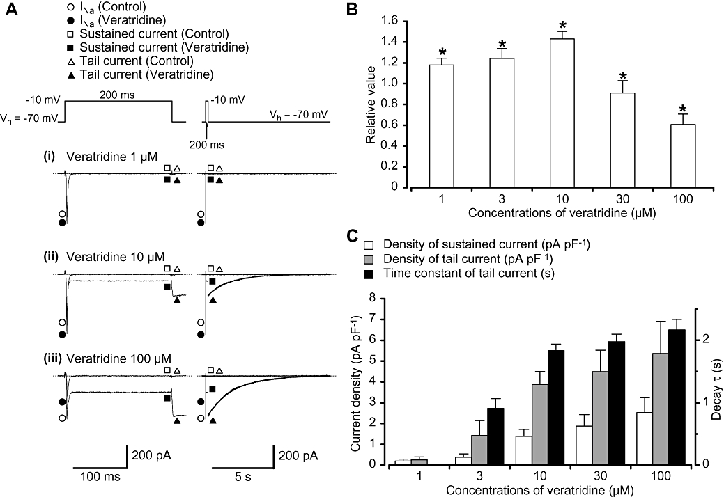
Effects of veratridine on the membrane currents in myocytes from mouse vas deferens using a conventional whole-cell configuration. (A) Representative current traces in the absence (control) and presence of veratridine (1 (i), 10 (ii) and 100 µM (iii)) using the same cell are shown at the left panel. The tail currents are also shown as an expanded trace at the right panel. The tail currents were well fitted by an exponential (10 µM, τ= 1.8 s; 100 µM, τ= 2.1 s). Membrane currents were evoked by a depolarizing rectangular pulse (200 ms duration; 20 s interval) from a holding potential of −70 to −10 mV. (B) Dual action of veratridine (1–100 µM) on the peak amplitude of INa. The relative values of the peak amplitude were expressed when the peak amplitude of INa in the absence of veratridine (control) was normalized as one. (C) Veratridine (1–100 µM) increased the amplitude of the sustained current (measuring the mean value of the last 20 ms of the test pulse) and the peak amplitude of the tail current in a concentration-dependent manner. The time constant of tail current decay (τ) was prolonged when the concentrations of veratridine were increased. The ordinate at the left side shows the current density of INa (pA pF−1). The time constant of the tail current decay (τ) was also indicated at the right side. Each column shows the mean of 5–17 cells (13 different animals) with +SD shown by vertical lines. Asterisk indicates a statistically significant difference and changes were considered significant at P < 0.05 (anova test).