
Figure 3.
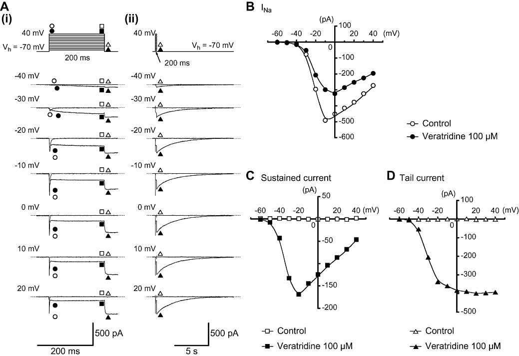
Effects of veratridine (100 µM) on INa in myocytes from mouse vas deferens. (A) (i) Original current traces were superimposed before (control) and after application of 100 µM veratridine at indicated pulse potentials. (ii) The decay of the tail current traces in the absence and presence of 100 µM veratridine superimposed at a slower time frame of (i). (B) Current–voltage relationships of the peak amplitude of transient INa obtained in the absence (control) or presence of 100 µM veratridine. The peak amplitude of INa was measured as the largest amplitude of the first 10 ms of the test pulse. (C and D) Current–voltage relationships of the 100 µM veratridine-induced sustained current (measured mean amplitude of the last 20 ms of the test pulse) and tail current (the peak amplitude). All curves were drawn by eye.