
Figure 4.
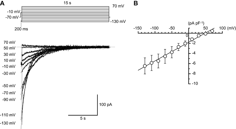
Ion selectivity of the veratridine-induced tail currents in myocytes from mouse vas deferens. (A) Current traces at the indicated membrane potentials. After an initial depolarization to −10 mV from a holding potential of −70 mV, the cell was repolarized to different test potentials (from −130 to 70 mV). (B) The peak amplitude of the veratridine-induced tail currents was plotted against the test potentials. The reversal potential was estimated to be 48.5 ± 1.6 mV.