
Figure 1.
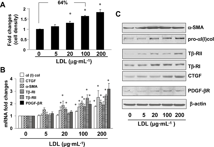
Low-density lipoprotein (LDL) stimulated HSC activation. Semi-confluent hepatic stellate cells (HSCs) were serum-starved for 24 h, followed by the addition of LDL at various concentrations as indicated for another 24 h. (A) MTS assays of cell proliferation. Results were expressed as cell density (%), compared with the untreated control [means ± standard deviation (SD)] (n= 3). *P < 0.05 versus cells with no treatment. (B) Real-time PCR analyses of the steady state levels of mRNA of genes relevant to HSC activation. The levels of target mRNAs were normalized with glyceraldehyde-3-phosphate dehydrogenase mRNA and were presented as mean ± SD (n= 3). *P < 0.05, compared with the untreated corresponding control (the first columns on the left side). (C) Western blotting analyses of proteins relevant to HSC activation. β-actin was used as an internal control for equal protein loading. Representative results from one of three independent experiments. α-SMA, α-smooth muscle actin; CTGF, connective tissue growth factor; PDGF-βR, platelet-derived growth factor-β receptor.