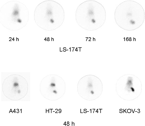
Figure 4.

γ-Scintigraphy of mice bearing tumour xenografts. Following i.v. injection with 80–100 µCi of 111In-CHX-A″-panitumumab, mice bearing LS-174T (upper row) xenografts were imaged over a 1 week period. γ-Scintigraphy was also conducted with other tumour xenograft models with representative scintigraphs taken at 48 h presented in the lower panel.
