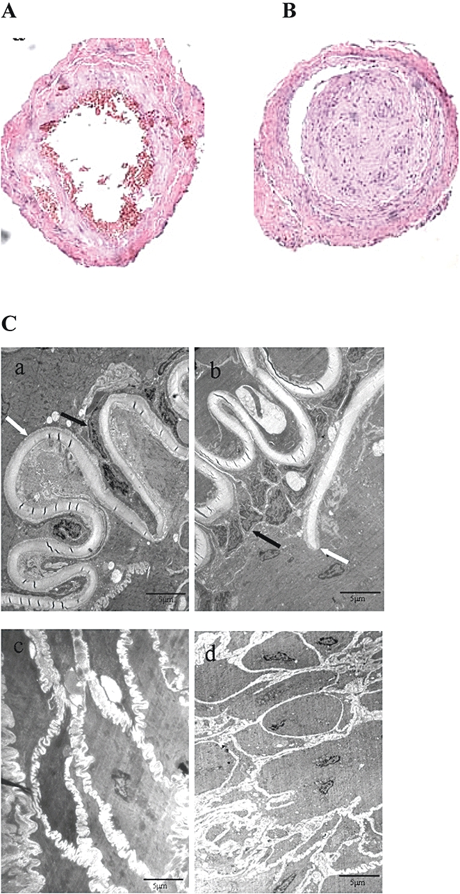
Figure 2.

Histological examination of femoral arteries in normal (A) and thromboangiitis obliterans (TAO) model (B) rats (haematoxylin and eosin staining, magnification: 100×). In (C), ultrastructure of femoral arteries in normal (a and c) and TAO model (b and d) rats. a and b, artery intima; c and d, medial membrane. Black arrows show endothelial cells and white arrows show inner elastic layer. In picture b, black arrow indicates endothelial cell proliferation, and the white arrow indicates discontinuity of the internal elastic lamina (transmission electron microscopy; magnification: 4000×).
