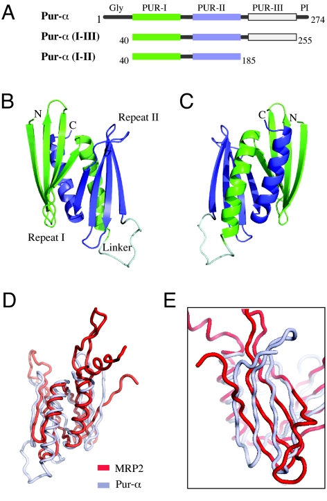
Fig. 1.
The structure of Drosophila Pur-α identifies the PUR domain as an MRP1/MRP2/P24-like nucleic-acid binding protein. (A) Schematic drawing of the distinct protein regions of Pur-α (Top) and the two protein fragments used in this study (Middle and Bottom). PUR repeats have been identified in this study by structure determination. (B) Ribbon backbone model of the globular domain formed by two PUR repeats, termed PUR domain. Similar to the schematic drawing in (A), PUR repeat I is shown in green, PUR repeat II in blue, and the linker connecting both repeats in gray. (C) View from (B) rotated by 180° around the vertical axis. (D) Superposition of the structure of Pur-α (gray) with the structure of MRP2 (red). Orientation is identical to (C). (E) Rotated and magnified superposition from (D) showing the orientation of the β-sheets from Pur-α and MRP2 with respect to each other.

