Fig. 3.
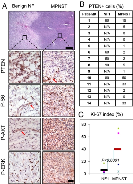
Decreased PTEN expression in human NF1-associated MPNSTs. (A) Histological and immunohistological analyses of transition zone of human NF1-MPNST lesions demonstrate reduced PTEN expression (second right panel; arrows indicates PTEN+ vascular endothelial cells) and activated PI3K and RAS/MAPK pathways. [Scale bars, 150 μm (H&E) and 25 μm (IHC).] (B) Summary of PTEN IHC staining results, presented as percentages of PTEN+ tumor cells, of 14 NF1 patients with MPNST lesions. N/A, only MPNST samples are available. (C) Human MPNST tumors have higher Ki-67 labeling index than NF tumors (P < 0.0001).

