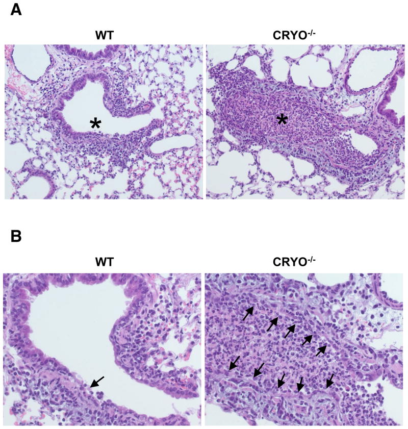
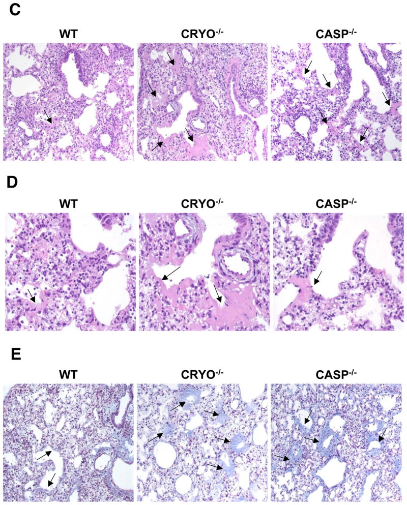
Figure 6. Virus-induced pulmonary necrosis is enhanced in the absence of cryopyrin.
Groups of 4 WT and cryopyrin−/− mice were infected i.n. with 8×10³ EID50 of PR8 influenza virus. Lung samples were collected on d3 and processed for routine H&E staining of 4μ paraffin sections. (A), top panels, ×200, WT airway epithelium is intact with only small foci of necrosis (arrow) and peribronchiolar inflammation; * shows unobstructed airway lumen. Cryopyrin−/−, the small bronchi and bronchioles show diffuse necrosis with loss of the airway epithelium and obstruction of the lumen by debris (*). (B), bottom panels ×400, Intact wildtype epithelium with limited foci of necrosis (arrow), and diffuse necrosis of CRYO−/− bronchiolar epithelium (arrows). Histologic evaluation was performed by an experienced veterinary pathologist (KLB). Groups of 4 wildtype and cryopyrin−/− mice were infected i.n. with 8×10³ EID50 of PR8 influenza virus. Lung samples were collected on d11 and 4μm paraffin sections processed for routine H&E (C,D) or Masson’s Trichrome staining (E). (C) ×200, the WT mice show only focal and minimal collagen deposition in the alveoli and occasionally in the terminal airways (arrows). This collagen deposition is exuberant in the mutant groups and often occludes alveoli and terminal airways (arrows). (D) ×400, arrows indicate areas of collagen deposition. (E) ×200, bottom row Masson’s Trichrome stain confirms collagen deposition. In the WT groups the insult was sufficiently severe to cause basement membrane damage, evidenced by a thin layer of collagen lining the alveolar spaces. In the mutant lungs, deposition of collagen is abundant and forms concentric layers that occlude alveoli and terminal airways.


