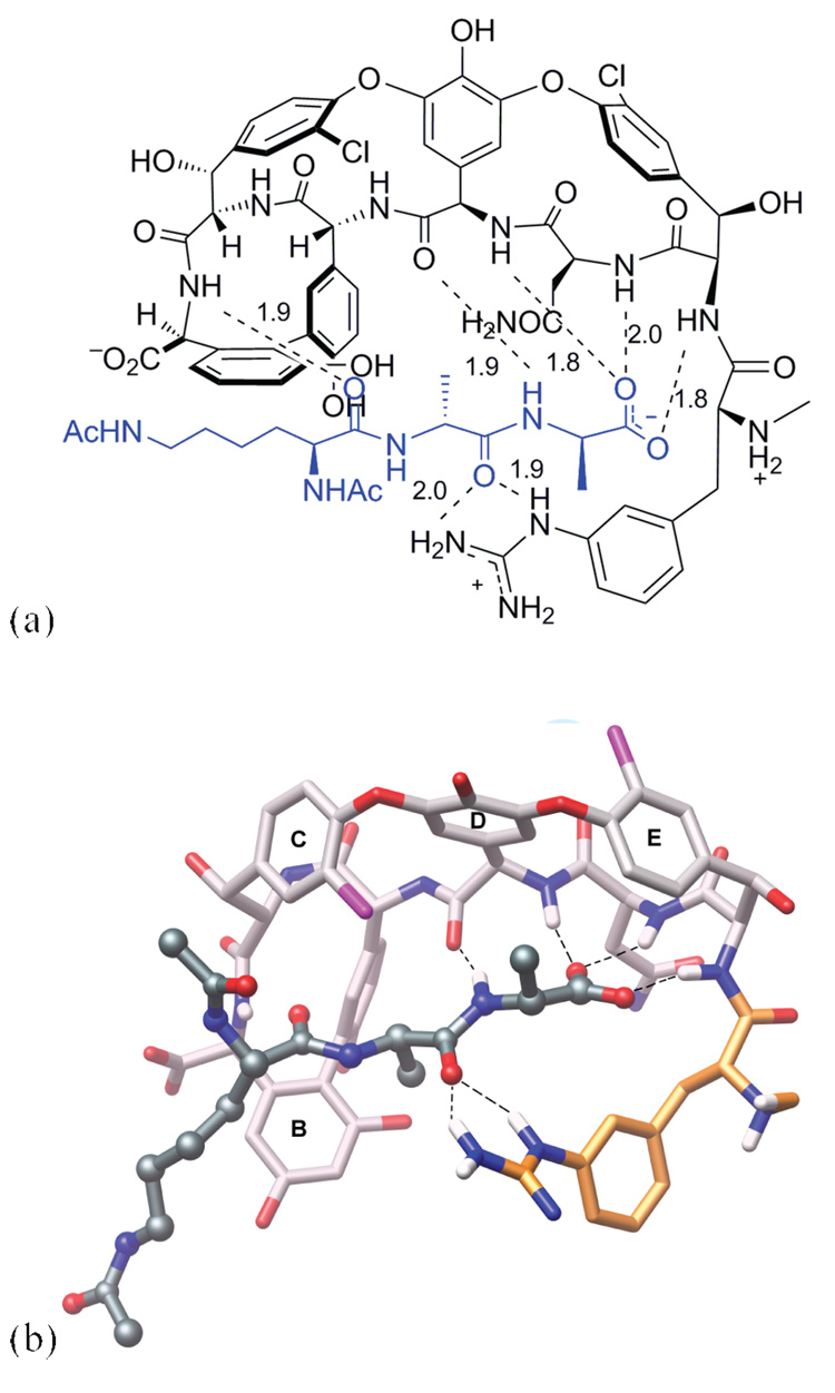
Figure 3.
Proposed binding of the N-terminally modified vancomycin analogue 11 with ligand 2 as determined by molecular modeling using the program MOLOC.7a,b Molecular modeling was preformed starting with the X-ray crystal structure of 5 co-crystallized with 2 determined to 1.8 Å-resolution (PDB Code: 1FVM).7c,d (a) Schematic view. (b) Ball and stick representation. Black dashed lines represent H-bonds. Distances between H-atom and heteroatom of H-bonding partner are given in angstroms. Color code: ligand skeleton: dark-grey; vancomycin skeleton: light-grey; N-terminal amino acid skeleton: orange; and O: red; N: blue; H: white. Figure was generated with the molecular graphics program Chimera.8

