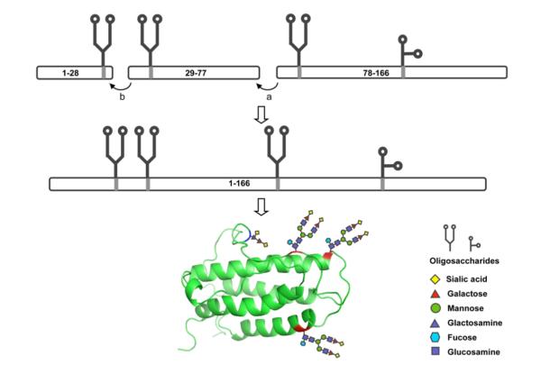
Figure 1.
Synthetic strategy toward homogeneous erythropoietin.
(a) Fragment condensation between G77 and Q78; (b) Native chemical ligation between G28 and C29. The ribbon diagram of the tertiary structure of human EPO protein is shown in green. N-linked glycosylation sites, N24, N38 and N83 are shown in red and O-linked glycosylation site, S126, in blue.

