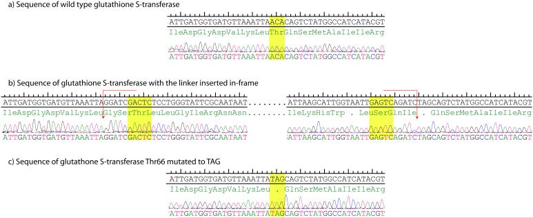
Figure 2.
Sequence of a single clone proceeding through the process. a. Sequence of wild-type glutathione S-transferase, the highlighted ACA is the codon removed by MlyI digestion of the transposon. b. The linker is inserted in-frame resulting in ampicillin resistance, the MlyI restriction sites are highlighted and red arrows indicate the cut sites. Upon digestion with MlyI and subsequent ligation the codon TAG (shown in yellow c.) replaces ACA.

