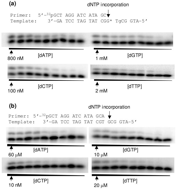
Figure 3.
Example gel images for the steady-state kinetic measurements for the nucleotide incorporation opposite the 8-oxodG portion of the 5′-Tg-(8-oxodG)-3′ tandem lesion (a) or the corresponding dG site for the undamaged substrate. (b). Klenow fragment (5 ng) was incubated with 10 nM DNA substrate at room temperature for 10 min. The highest dNTP concentration is shown in the figure, and the ratio of dNTP concentrations between adjacent lanes was 0.5-0.6.

