
Figure 4.
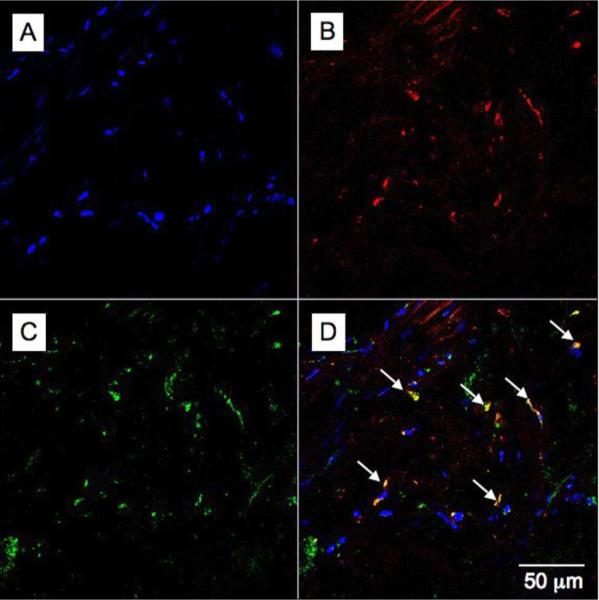
Confocal microscopy images of aortic tissue excised from an apoE-KO mouse injected with the 18A-Gd agent 24 hours prior to excision. A - DAPI staining for nuclei (blue), B - the rhodamine channel (red) indicating areas of 18A-Gd uptake, C - Alexa 647:CD68 staining for macrophages (green) and D - merging the previous 3 images reveals the affinity of agent 18A-Gd for macrophages (yellow).