
Figure 2. While all isolated cells load with Fura-2, only SCCs display GFP fluorescence.
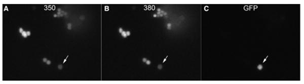
A. All isolated epithelial cells show Fura-2 fluorescence at 350 nm. B. Fura-2 fluorescence is detected in all isolated cells at 380 nm. C. Only SCCs express GFP fluorescence (arrow). Images taken with at 40× oil immersion lens.