
Figure 8.
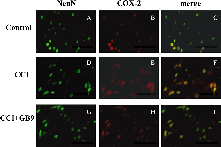
Double-labelled immunofluorescent staining of NeuN (green) and cyclooxygenase-2 (COX-2) (red) in the dorsal region of the lumbar spinal cord ipsilateral to the injury after i.p. GB9 (10 mg·kg−1, i.p.) administration, showing spinal cord sections from the control (A–C), chronic constriction injury (CCI) (D–F), and CCI + GB9 (G–I) groups. The images represent multiple fields examined for each group from three independent immunofluorescence observations. The immunostaining images show cells labelled with NeuN (green) and COX-2 (red) in the spinal cord. The merged images of C, F and I (yellow) indicate colocalization of COX-2 and NeuN (neuronal specific marker) immunoreactive cells in the spinal cord. The results of double immunofluorescent staining from the control, CCI and CCI + GB9 groups all showed that COX-2 was colocalized with NeuN (C, F and I). COX-2 was stronger in the CCI group than in the control and CCI + GB9 groups. Scale bars: 50 µm for all images.