Figure 2.
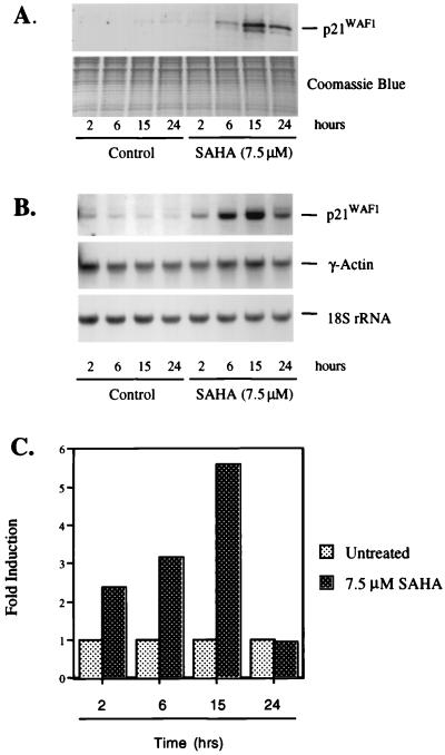
SAHA induces p21WAF1 mRNA and protein in T24 cells. T24 cells were cultured with SAHA (7.5 μM) for the indicated times. (A) Western blot analysis of p21WAF1 protein expression. Protein extracts were prepared and resolved (25 μg) on 15% SDS/PAGE. p21WAF1 protein was detected by using the mouse monoclonal anti-p21WAF1 F5 antibody. A parallel gel was stained with Coomassie blue for a loading control. (B) Northern blot analysis of p21WAF1 mRNA expression. Total RNA (10 μg) was isolated and fractionated on 1.2% agarose and transferred to a nylon membrane. The membrane was probed by using 32P-random-labeled human p21WAF1 cDNA, the γ-actin cDNA, and a 50-mer oligonucleotide specific for 18S rRNA. (C) Run-on transcription assay by using nuclei from SAHA-induced T24 cells. Nuclei were prepared and labeled with [α32P]-CTP. Equal levels of radioactivity (approximately 5 × 10⁷ cpm) were incubated with filters containing 5 μg of the indicated plasmid cDNA. Radioactivity bound to the filters was quantified and normalized to actin at each time point examined. The actin-normalized value for the untreated sample was adjusted to a value of 1 to determine fold increase after culture with SAHA.

