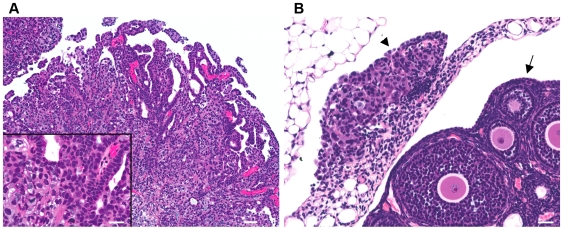
Figure 2. Human OVCA cell xenograft in mouse peritoneal cavity.
(A) Representative OVCA exhibits predominantly solid tumor growth, with evidence of papillary projections and occasional minute cysts (bar = 25 µm). Inset, higher resolution photomicrograph depicting cuboidal to polygonal pleomorphic microcystic epithelium with anisokaryosis and atypical mitoses of ovarian cancer cell nuclei (bar = 50 µm). (B) OVCA tumor nodule implant on uterine infundibulo-ovarian ligament (arrowhead) adjacent to the ovary (arrow) (bar = 25 µm). Tumor implants occurred throughout the peritoneal cavity, peri-ovarian connective tissues, and invaded surrounding tissues such as intestines, liver and diaphragm. Hematoxylin and eosin (H&E) stained, paraffin-embedded tissue sections.

