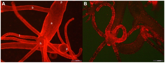
Figure 1. In vivo fluorescence imaging of Hydra vulgaris exposed to QRs for different times.
A) In vivo image of two Hydra, 30 minutes p.i.: QR red fluorescence labels uniformly all body regions, from the tentacles (t) to the peduncle (p) located in the upper part of the image. In this picture an adult (a) with a bud (b) on the left side turns towards the camera the hypostome (h), surrounded by a ring of tentacles. A second Hydra is placed horizontally below, bending the peduncle round the camera. B) In vivo image of different polyps, 2 h p.i. with QRs. In the foreground a Hydra turns the hypostome toward the bottom. A strong punctuated fluorescence labels the mouth, the tentacles and at a lower extent the animal body. Scale bar 500 µm.

