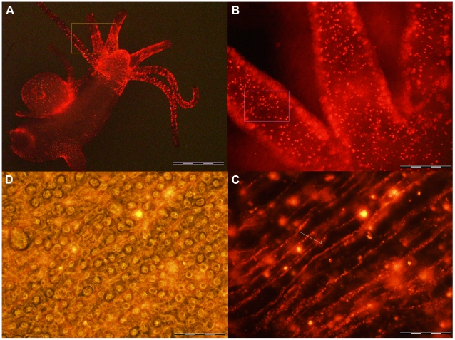
Figure 2. Tissue distribution of QR labelled cells.
A) In vivo imaging of a whole animal, incubated with QRs in SolHy, at pH 4, for 2 h, washed and observed by fluorescence microscopy. QR staining is distributed on tentacles, hypostome and peduncle. The region within the orange frame is shown at higher magnification in B) punctuated fluorescence is uniformly distributed over the tentacles. Regions out of focus belong to different planar levels. C) Image showing a region within the pink inset of B, at higher magnification. Single animals were put on a microscope concave glass slide, with a drop of SolHy and observed under fluorescence or phase contrast mode (D). QRs label cell membranes of battery cells (arrows), but are also compacted into cytoplasmic granule-like structures, which do not colocalize with nematocytes, as evident in the bright field-fluorescence merged image (D). The round structures represent the numerous nematocytes (desmonemes, stenotheles and izorhiza) embedded in the cytoplasm of the battery cells. Scale bars: 1 mm in A, 200 µm in B, 20 µm in C e D.

