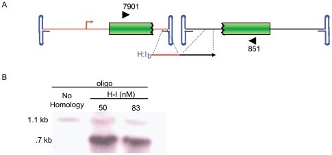
Figure 4. Junction Characterization of Functional Concatemers.
A) This cartoon depicts our AAV split gfp vector system and the position of primers (block arrows) used for functional concatemer amplification. B. Evaluation of amplification products by southern blotting (gfp probe) showed a unique band formed only from DNA isolated for H-I treated cells. This band was directly cloned and sequenced.

