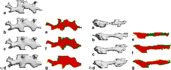
Fig. 7.

A trabecula in two PTH-treated ovariectomized rats was tracked over time to determine the development of bone formation (1 and 2). On the left of 1 and 2, you see three-dimensional segmented images of a trabecula, after PTH treatment is started at week 8, taken at weeks 8 (a), 10 (b), 12 (c), and 14 (d). On the right, you see overlaid two-dimensional segmented sections comparing weeks 8 and 10 (e), 10 and 12 (f), and 12 and 14 (g). Yellow indicates resorbed bone, green newly formed bone, and red unchanged bone. Bone formation is clearly seen over time in both trabeculae. In trabecula 1, bone is mostly deposited in the cavities in the first 2 weeks, while later on bone is added to the surface. In trabecula 2, the trabecula appears cleaved after segmentation, although most likely there was still a thin line of bone present. PTH treatment leads to bone formation at the cleaved site, where it is most needed hereby restoring the trabecula
