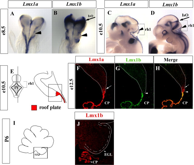
Figure 1.
Expression of Lmx1a and Lmx1b in rh1 roof plate and its derivative, the choroid plexus epithelium. A–D, Whole-mount in situ hybridization using probes for Lmx1a and Lmx1b. A, B, Dorsal views of E8.5 embryos. Lmx1a and Lmx1b were expressed in roof plate progenitor cells at the lateral edges of the neural tube (arrowheads). Lmx1b was also expressed across the isthmic organizer (IsO) at the mid/hindbrain junction (arrow in B). C, D, Lateral view of E10.5 embryos. Brackets indicate rhombomere 1 (rh1). C, Lmx1a was expressed in the roof plate along the anterior–posterior axis of the embryo, including rh1 (arrowhead). D, Lmx1b was expressed in the anterior roof plate (arrowhead) and in the IsO (arrow). E, Schematic of dorsal view of rh1 and paramedial sagittal section through rh1 at E10.5. Boxed region is equivalent to sagittal sections in F–H. F, Immunohistochemistry at E12.5 showed expression of Lmx1a in choroid plexus (CP) and in the rhombic lip (RL) (arrow). G, H, Arrowhead shows the CP/RL boundary. G, Lmx1b expression was restricted to the CP. H, Merged image of F and G. I, Schematic of midsagittal section of P6 cerebellum. Boxed region identifies the posterior lobe and the CP shown in the next panel. J, Lmx1b was expressed in the CP but not in the cerebellum. EGL, External granule layer; IV, Fourth ventricle.

