Figure 6.
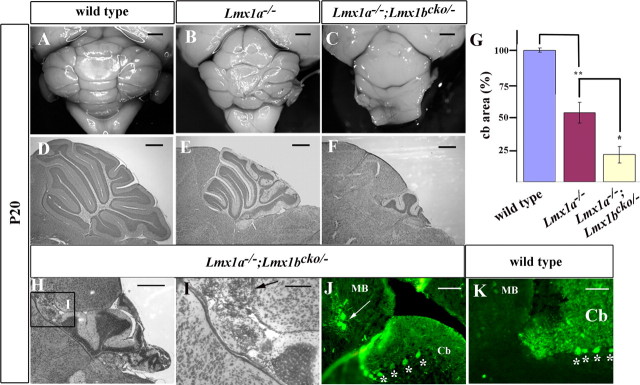
Cerebellar morphogenesis is dependent on Lmx1-dependent roof plate signaling. A–F, Whole mount (A–C) and Nissl-stained midsagittal sections of P20 cerebella (D–F) demonstrating that loss of both Lmx1b and Lmx1a had significant effects on cerebellar development. The cerebella of Lmx1a−/− mice were small, but those of Lmx1a−/−; Lmx1bcko/− mice were even smaller. G, Quantification of midsagittal sections of P20 cerebella (n = 4). Area of wild type is indicated as 100%. *p < 0.01; **p < 0.001. Error bars indicate SE. H–J, Ectopic cerebellar cells were observed in n = 2/8 Lmx1a−/−; Lmx1bcko/− cerebella in the midbrain, evident in Nissl-stained sections. I, Higher-magnification of the boxed region in H showing ectopic cerebellar cells in the midbrain. J, White arrows indicate calbindin-positive Purkinje cells present in the midbrain (MB), and cerebellar (Cb) Purkinje cells are indicated by *, in similar sections. K, An equivalent wild-type calbindin-stained section. Scale bars: A–C, 1 mm; D–F, 500 μm; H, 500 μm; I–K, 250 μm.

