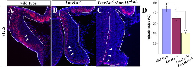
Figure 7.
Reduced proliferation in the cerebellar anlage correlates with reduced roof plate size. A–C, Midsagittal sections of BrdU (red) and DAPI (blue)-stained wild-type (A), Lmx1a−/− (B), and Lmx1a−/−; Lmx1bcko/− (C) cerebella anlage at E12.5. Arrowheads show BrdU staining in the ventricular zone. Proliferation was reduced in Lmx1a−/− and Lmx1a−/−; Lmx1bcko/− embryos. D, Mitotic index of E12.5 ventricular zone (BrdU/DAPI). Lmx1a−/−; Lmx1bcko/− embryos showed significant reduction compared with Lmx1a−/− embryos, which was reduced compared with wild-type embryos. *p < 0.01; **p < 0.001. Error bars indicate SE.

